

Testing for CKD

Kidney function: glomerular filtration rate (eGFR test)
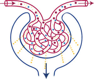
Normal Filtration

Diagram: The kidneys contain around a million tiny filtering units called nephrons. Each nephron (shown) has a cluster of blood vessels called the glomerulus, where filtering happens. Blood flows into the glomerulus through one small vessel and out through another. As blood passes through, water and small waste products (yellow dots) pass out of the blood vessels and into a surrounding cup-like structure called Bowman’s capsule. This fluid is now called filtrate, and it goes on to become urine. GFR is the amount of filtrate produced per minute, added up across all the nephrons in both kidneys.
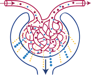

Kidney damage: albuminuria/proteinuria (uACR test)

glomerulus proteinuria

Diagram: When the kidneys are functioning normally, larger particles such as blood cells and proteins (red and blue dots) are too large to pass through the glomerular filter and remain in the bloodstream. Damage to the glomeruli, for example from poorly controlled diabetes or hypertension, increases the permeability of the filter, allowing proteins (blue dots) and sometimes blood cells (red dots) to leak into the filtrate and eventually into the urine. The amount of albumin leaking into the urine is measured using uACR. Higher uACR values indicate greater glomerular damage.

Assess:

  • Estimated glomerular filtration rate (eGFR) – to measure kidney function, using the CKD-EPI (2009) formula
  • Urinary albumin: creatinine ration (uACR) – to detect kidney damage
  • Urine dip – to rule out acute urinary tract infection/ incidental finding of haematuria

Identify aetiology where possible: hypertension, diabetes, polycystic kidneys, glomerulonephritis, acute on chronic kidney disease.

heart icon
Why use both uACR and eGFR?

eGFR measures kidney function; uACR measures kidney damage. uACR may detect CKD earlier than eGFR, particularly in people with diabetes or hypertension. A persistent uACR ≥3 mg/mmol indicates CKD even when eGFR is normal.

Albuminuria is an independent risk factor for both CKD progression and cardiovascular disease. Using uACR and eGFR together predicts risk more accurately than either alone, and guides monitoring frequency, treatment and referral.

Further parameters to aid diagnosis:

  • Urinalysis – to detect presence of persistent haematuria
  • HbA1c – to diagnose/ exclude diabetes or assess control if known diabetic.
  • FBC if eGFR <45 ml/min/1.73m² to rule out renal anaemia.
  • Myeloma screen (if not done previously or cause is unclear) – serum electrophoresis, Bence-Jones protein, serum free light chains
  • Renal ultrasound – not routinely required except for:
    • Accelerated progression of CKD (decline in eGFR ≥ 25% in 1 year OR ≥ 15 ml/min/1.73m² in 1 year)
    • Visible or persistent invisible haematuria
    • Symptoms suggestive of urinary obstruction (urinary retention/decreased or altered flow/ hesitancy/ loin pain/ increased urgency and nocturia/ incontinence)
    • Family history of genetic renal disease e.g. ADPKD
    • Consider if new diagnosis of CKD stages G4 or G5 (not known to nephrology)

warning icon
Presence of haematuria
  • Visible (macroscopic/ frank) haematuria:
    • Usually fast track urology referral for imaging and cystoscopy unless strong pointers to acute renal disease.
  • Non-visible (microscopic) haematuria:
    • Use reagent strip to detect.
    • Non-visible haematuria should be confirmed on at least 2 out of 3 consecutive urine tests (taken on separate occasions).
    • Evaluate further if there is a result of 1+ or more.
    • Measure renal function and assess for proteinuria in all cases.
    • Management:
      • Age >60:Usually refer to urology (see local guidance for referral pathways). If renal impairment and/or proteinuria also present, consider simultaneous referral to nephrology.
      • Age <60 or >60 with negative urological investigations: If no proteinuria AND eGFR > 60 ml/min/1.73m² manage as CKD stage G1/G2 otherwise refer to nephrology.

More information about testing

Kidney assessment includes tests of both kidney function (eGFR, typically, and CrCl, for higher risk drugs or extremes of muscle mass) and tests for kidney damage, which look for protein in the urine. Use the accordions below to learn more.

Estimated glomerular filtration rate

Glomerular Filtration Rate (GFR) is the standard measure of kidney function. Measurement of true GFR is not practical in routine clinical practice as it is time consuming and expensive. Therefore, standardised formulae have been developed to calculate estimated GFR (eGFR). These formulae incorporate serum or plasma creatinine, age and sex. eGFR is routinely calculated by all laboratories in the United Kingdom and reported alongside creatinine values.

Current NICE guidance recommends use of the ‘Chronic Kidney Disease Epidemiology Collaboration (CKD-EPI) (2009)’ equation for this purpose.

  • NICE recommends that this equation is used without an ethnicity adjustment factor.
  • The CKD-EPI 2021 equation should not be used as it has not been validated in UK populations.

Creatinine Clearance (Cockroft & Gault)

In product literature for therapeutic drugs, the effects of kidney impairment on drug elimination are usually stated in terms of creatinine clearance (CrCl) as a surrogate measure for GFR. Although eGFR and CrCl are not interchangeable, eGFR can be used to guide dose adjustment for most medicines in most adults of average body size.

CrCl should be used in preference to eGFR in situations where greater accuracy is required, including for toxic drugs, drugs with a narrow therapeutic index that are primarily renally excreted, older adults, and patients with extremes of muscle mass. In addition, the Medicines and Healthcare products Regulatory Agency (MHRA) advises that CrCl should be used to estimate renal function when dosing direct-acting oral anticoagulants (DOACs).

Albuminuria/ Proteinuria

Excessive albumin/protein excretion in the urine is a significant risk factor for both CKD progression and cardiovascular morbidity and mortality. Unlike haematuria, albuminuria is almost always kidney in origin. In clinical practice, albuminuria is measured using a spot urine sample to calculate the urinary albumin-to-creatinine ratio (uACR).

  • Urinary albumin leak may be the very first sign of kidney disease for many patients, such as those with diabetic kidney disease, and may appear many years before eGFR starts to decline.
  • Albuminuria is an important independent risk factor for cardiovascular morbidity and mortality.
  • Measurement of uACR forms part of the NICE classification system for CKD.
  • In people with CKD, quantification of albuminuria allows risk-stratification and identifies those at greatest risk of progression to kidney failure.
  • The level of albuminuria will help determine optimum patient management e.g. frequency of monitoring, blood pressure target, eligibility for disease modifying therapies such as renin-angiotensin-aldosterone-system (RAAS) inhibitors and sodium-glucose co-transporter-2 (SGLT2) inhibitors, alongside timing of referral to see a specialist service (see pharmacological management section).
  • Excessive albuminuria, or albuminuria accompanied by haematuria, may indicate an underlying glomerular disorder, particularly in patients with other features of autoimmune or multisystem disease. These patients should be considered for urgent specialist assessment (see referral section). As a guide, people without diabetes should be referred for nephrology evaluation if they have:
    • ACR ≥70 mg/mmol or
    • ACR ≥30 mg/mmol with haematuria (confirmed on urinary dipstick testing).
  • In those with albuminuria, the risk of kidney failure is greater in younger people and the risk of cardiovascular disease is greater in older people. These risks may be altered by therapy.